November 18, 2021
Researchers study the link between vitamin D and inflammation

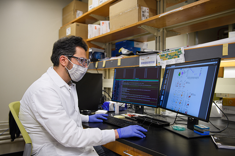
Majid Kazemian and a team of scientists have discovered that a form of vitamin D (not the over-the-counter pills) could help combat the inflammation in cells of people with severe cases of COVID-19. (Purdue University photo/Rebecca McElhoe.)
An active metabolite of vitamin D—(not the over-the-counter version) — is involved in shutting down inflammation, which could potentially be beneficial in patients with severe COVID-19.
WEST LAFAYETTE, Ind. — Scientists recently gained insights into how vitamin D functions to reduce inflammation caused by immune cells that might be relevant to the responses during severe COVID-19. In a study jointly published by Purdue University and the National Institutes of Health, scientists do just that.
Majid Kazemian, assistant professor in the departments of Computer Science and Biochemistry at Purdue University, was co-lead author of the highly collaborative study, along with Dr. Behdad Afzali, chief of the Immunoregulation Section of the National Institutes of Health’s National Institute of Diabetes and Digestive and Kidney Diseases.
“Our work demonstrates a mechanism by which vitamin D reduces inflammation caused by T cells. These are important cells of the immune system and implicated as part of the immune response to the infection causing COVID-19. Further research, especially clinical trials, and testing in patients, are necessary before this can be adopted as a treatment option.” Kazemian said. “We do not recommend the use of normal vitamin D off the shelf at the pharmacy. No one should be taking more than the recommended doses of vitamin D in an attempt to prevent or combat COVID infections.”
Previous studies have shown vitamin D’s ability to reduce the inflammation caused by T cells — inflamed cells in the lung characteristic of the most severe and dangerous cases of COVID-19. But as important as understanding that a drug works is understanding the how and the why. This is both to maximize benefit and minimize harm (such as preventing people from eating livestock dewormer or injecting household cleaners into their veins) as well as to pave the way for future treatments.
If scientists understand how vitamin D works to combat inflammation, they understand more about how both the drug and related diseases work, paving the way for new, even more effective drugs.
Kazemian and his team began by studying how viruses affect lung cells in a previous study. Finding that viruses can trigger a biochemical pathway, known as the immune complement system, the researchers started looking for ways to disrupt that pathway and ameliorate the subsequent inflammation.
The team studied and analyzed individual lung cells from eight people with COVID — something only possible because of Kazemian’s experience with gene sequencing and data mining. They found that in the lung cells of people with COVID, part of the immune response was going into overdrive, exacerbating lung inflammation.
“In normal infections, Th1 cells, a subset of T cells, go through a pro-inflammatory phase,” Kazemian said. “The pro-inflammatory phase clears the infection, and then the system shuts down and goes to anti-inflammatory phase. Vitamin D helps to speed up this transition from pro-inflammatory to the anti-inflammatory phase of the T cells. We don’t know definitively, but theorize the vitamin could potentially help patients with severe inflammation caused by Th1 cells.”
In patients with COVID-19, the pro-inflammatory phase of the Th1 cells seems not switched off, possibly because the patients didn’t have enough vitamin D in their system or because something about the cell’s response to vitamin D was abnormal. In that case, the researchers posit, adding vitamin D to existing treatments in the form of a prescribed highly concentrated intravenous metabolite may further help people recovery from COVID infections, though they have not tested this theory.
“We found that vitamin D – a specialized form of it, not the form you can get at the drugstore — has the potential to reduce inflammation in the test tube, and we figured out how and why it does that,” Kazemian said. However, it’s important to understand that we did not carry out a clinical study, and the results of our experiments in the test tube need to be tested in clinical trials in actual patients.”
The work was funded by NIGMS, NIDDK, NIAID and NHLBI of the NIH, with additional funding from the Wellcome Trust, the Crohn’s and Colitis Foundation of America, the British Heart Foundation, the Showalter Trust, the German Research Foundation, the National Agency of Research and Development of Chile, and England’s National Health Service’s National Institute for Health Research Biomedical Research Centre and Research Facility.
About Purdue University
Purdue University is a top public research institution developing practical solutions to today’s toughest challenges. Ranked in each of the last four years as one of the 10 Most Innovative universities in the United States by U.S. News & World Report, Purdue delivers world-changing research and out-of-this-world discovery. Committed to hands-on and online, real-world learning, Purdue offers a transformative education to all. Committed to affordability and accessibility, Purdue has frozen tuition and most fees at 2012-13 levels, enabling more students than ever to graduate debt-free. See how Purdue never stops in the persistent pursuit of the next giant leap at https://purdue.edu/.
Writer, Media contact: Brittany Steff, bsteff@purdue.edu
Source: Majid Kazemian, kazemian@purdue.edu
ABSTRACT
Autocrine Vitamin D-signaling switches off pro-inflammatory programs of Th1 cells
Daniel Chauss, Tilo Freiwald, Reuben McGregor, Bingyu Yan, Luopin Wang, Estefania Nova-Lamperti, Dhaneshwar Kumar, Zonghao Zhang, Heather Teague, Erin E West, Kevin M Vannella, Marcos J Ramos-Benitez, Jack Bibby, Audrey Kelly, Amna Malik, Alexandra F Freeman, Daniella M Schwartz, Didier Portilla, Daniel S Chertow, Susan John, Paul Lavender, Claudia Kemper, Giovanna Lombardi, Nehal N Mehta, Nichola Cooper, Michail S Lionakis, Arian Laurence, Majid Kazemian, Behdad Afzali
DOI : 10.1038/s41590-021-01080-3
The molecular mechanisms governing orderly shutdown and retraction of CD4+ T helper (Th)1 responses remain poorly understood. Here, we show that complement triggers contraction of Th1 responses by inducing intrinsic expression of the vitamin D (VitD) receptor (VDR) and the VitD-activating enzyme CYP27B1, permitting T cells to both activate and respond to VitD. VitD then initiated transition from pro-inflammatory IFN-g + Th1 cells to suppressive IL-10+ cells. This process was primed by dynamic changes in the epigenetic landscape of CD4+ T cells, generating super-enhancers and recruiting several transcription factors, notably c-JUN, STAT3 and BACH2, which together with VDR shaped the transcriptional response to VitD. Accordingly, VitD did not induce IL-10 in cells with dysfunctional BACH2 or STAT3. Bronchoalveolar lavage fluid CD4+ T cells of COVID-19 patients were Th1-skewed and showed de-repression of genes down-regulated by VitD, either from lack of substrate (VitD deficiency) and/or abnormal regulation of this system.
Note to journalists: Journalists visiting campus should follow visitor health guidelines.

